Makler counting chamber for motility and concentration analysis
Makler counting chamber for motility and concentration analysis
Click image to enlarge
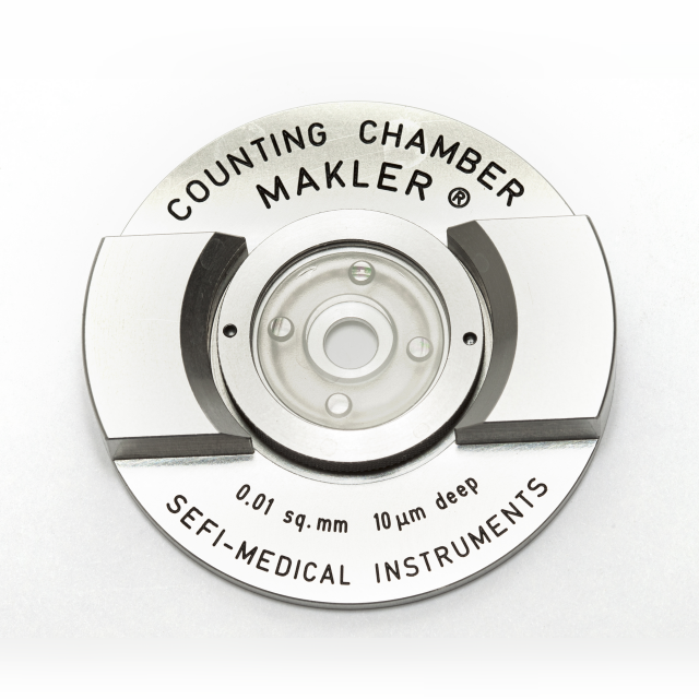
The Makler counting chamber is a simple-to-use device for rapid and accurate sperm count, motility and morphology evaluation, from undiluted specimen.
The chamber is composed of two parts:
1. The lower part has a metal base and two handles. In the center of the base there is a flat disc made of optical flat glass on which the sample is placed.
2. The upper part is the cover glass encircled with a metal ring. At the center of its lower surface there is a 1mm2 grid, subdivided into 100 squares, each one of 0.1 x 0.1 mm.
When the cover glass is placed on the four tips, the space bounded in a row of 10 squares is exactly one millionth of mL. Therefore, the number of sperm heads in 10 squares indicates their concentration in million/mL.
More information:
Unique features and advantages:
Easy to use:Sperm count performed from undiluted specimen. Fast results:
The number of spermatozoa counted in any strip of 10 squares of the grid indicates their concentration in millions/mL. No additional factors are necessary for calculation. Optimal depth:
The depth of 10 microns eliminates blurring and allows sperm to move freely. The applied sample is observed in one focal plane. Built-in grid:
The grid is on the cover glass. This eliminates the need to insert a grid into the microscope eye piece and remove it when not required. Economical:
Reusable. Easily cleaned with a non-bleach disinfectant solution. Self-controlled accuracy:
Observation of color fringes at the four contact points, provides a self-controlled test for accuracy. The cover glass can never be raised by the applied sample. Calibration unnecessary:
Repeated use with complete accuracy without calibration. Superior technology:
Manufactured by state-of-the-art precision engineering. Checked individually by laser beam for precision and accuracy.
Accessories:
Cleaning brush. Lint free lens paper. Chamber grip – this device should be placed on the stage of the microscope during sperm analysis. It grips the chamber tightly and enables smooth shifting.Additional information:
| Reference: MKLR |
| Catalog: Download catalog |
| Protocol: Download protocol |