Importance of microscopy for cell biologists in general and spermatologists specifically
Part 2: Specialized microscopic techniques for sperm
In the second blog on Microscopy, I indicated that brightfield microscopy, positive and negative phase microscopy and fluorescence microscopy are now routinely used for CASA analysis. Before proceeding to a more detailed discussion of fluorescence microscopy I will first discuss a complimentary technique to phase contrast microscopy namely Nomarski Differential Interference Microscopy.
1. Nomarski Differential Interference Microscopy (NDIC)
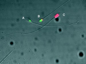
It is a contrast enhancing technique that allows visualization of live cells without staining like phase contrast. However, Wollaston prisms are utilized that assist to provide remarkable sharp and three dimensional like images. It is particularly useful to study human and animal sperm. Moreover, it has been very extensively used in the field of IVF for egg and sperm manipulation or similar optics, called Hoffman optics. NDIC has a lot of potential in CASA but unfortunately is very expensive for routine CASA. In Fig. 1 sperm of two ferret species could be distinguished on the basis of the structure of the posterior border of the acrosome.
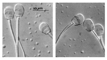
Fig. 1: NDIC images of domestic ferret on the left and black footed ferret sperm on the right (van der Horst et al. 1991).
2. Fluorescence microscopy:
Three essential components of epifluorescence is a high intensity light source, a special cube (Fig. 2) controlling specific incoming and very specific emitting fluorescent wavelengths and special objective quarts lenses. Nikon in their webpage states: “The technique of fluorescence microscopy has become an essential tool in biology and the biomedical sciences, due to attributes that are not readily available with traditional optical microscopy. The application of an array of fluorochromes has made it possible to identify cells and sub-microscopic cellular components with a high degree of specificity. In fact, the fluorescence microscope is capable of revealing the presence of a single molecule. Through the use of multiple fluorescence labeling, different probes can simultaneously identify several target molecules simultaneously”. It is potentially possible to determine almost any structure of a sperm cell such as the acrosome which is lectin specific (Fig. 3) or inorganic chemical compounds such as Calcium or various functional proteins and even molecules and in several cases multiple probes can be used.
-
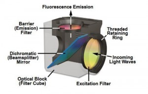
- Fig. 2: Typical filter cube (from Nikon webpage) used in a Nikon fluorescent microscope showing the different filters. For a specific fluorochrome a cube is selected that has a very specific excitation filter only allowing very specific wavelengths through, a dichroic mirror and a barrier filter which then narrows the wavelength that will reach the camera or eye.
In Fig. 4 two labels are used simultaneously to show in red (Mitotracker red) that the mitochondrial membrane potential is intact and in blue (Hoechst) the sperm head with intact nucleus.
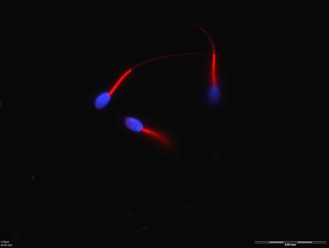
Fig. 4: Sperm of Rhesus monkeys showing in red the midpiece with mitochondria showing active mitochondrial membrane potential and blue head with intact chromatin of nucleus (van der Horst and Maree, 2014).
3. Laser Confocal microscopy
Laser Confocal microscopy offers the remarkable ability of depth of focus, elimination of out-of-focus glare, and the ability to produce serial optical sections from thick specimens which allows remarkable 3-D reconstruction of cells. One of the reasons is of course the much shorter wavelength of laser compared to light and therefore with laser better resolution can be obtained of the specimens. With confocal microscopy imaging of either fixed or living sperm usually been labeled with one or more fluorescent probes could be performed (Fig. 5B). A remarkable facet allows following the production and movement of proteins in cells. Fig. 5A is a diagram showing the complexity of the basic system.
-
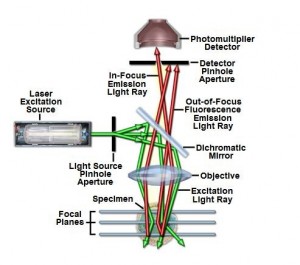
- Fig. 5A: Configuration of Laser confocal microscopy.
-
- Fig. 5B: Lipid peroxidation in stallion sperm laser confocal microscopy (C Ortega Ferrusola et al 2009- Reproduction).
4. Scanning (SEM) and Transmission (TEM) electron microscopy
Essentially in these microscopes electrons are accelerated in a vacuum and use electro magnetic lenses to focus the electron beam on the image and electro magnetic objective and projective lenses will enlarge and project the image. In principle the resolution of the transmission electron microscope is x1000 more than in a light microscope. Very specialized preparation procedures are required to produce a usable specimen. Scanning electron microscopy provide detail and chemical composition of the surface of cells and transmission electron microscopy provide detail of the organelles of the cell.
Fig. 6 represents SEM of sperm of four ferret species and show detail of the posterior border of the acrosome. Fig. 7 shows the transmission electron microscopy of the head and midpiece of African elephant sperm.
-
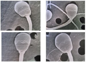
- Fig. 6: SEM of four ferret species van der Horst (1991).
-
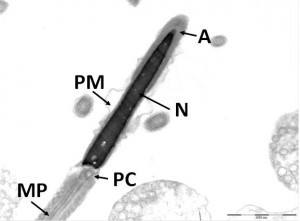
- Fig. 7: TEM of African elephant sperm showing nucleus, acrosome and midpiece with mitochondria (Luther 2017).
Summary: Apart from bright field, phase contrast microscopy and Fluorecence microscopy typically used for CASA, there is a vast range of other microscopic techniques to study sperm. I have only touched on specialized microscopy and there are many others such as atomic force microscopy. However, I hope that three blogs on microscopy serve to stimulate users of CASA to realize the need to study sperm also using these amazing complimentary techniques.
The End
Gerhard van der Horst (PhD, PhD)
Senior Consultant
MICROPTIC S.L.